KOKorean
 ENEnglish
 JP日本語
 CN中文(简体)
 TW中文(繁體)


Recurrent Sebaceous Cyst in Seoul: Why It Comes Back and What Removing It Again Can and Cannot Do
A recurrent sebaceous cyst is the same lump returning where a cyst was squeezed or removed, and the most common cause is a sac wall left behind. The lining of that wall keeps making keratin, so squeezing out the contents or draining pus tends to let it refill. Removing the wall lowers the chance of recurrence, but surgery does not mean it can never return: a thin wall that tears, or a wall stuck in scar tissue from earlier inflammation, can leave a small piece behind, and new cysts elsewhere cannot be prevented. How likely recurrence is depends on the site and on earlier treatment. Bonita Clinic does not squeeze cysts but removes the sac wall as a rule, and a recurrent cyst is examined for inflammation and scar tissue before the method is chosen. If you are planning treatment in Seoul, you can send photos of the lump and the old scar first.
Curious about your own case? Ask us online →
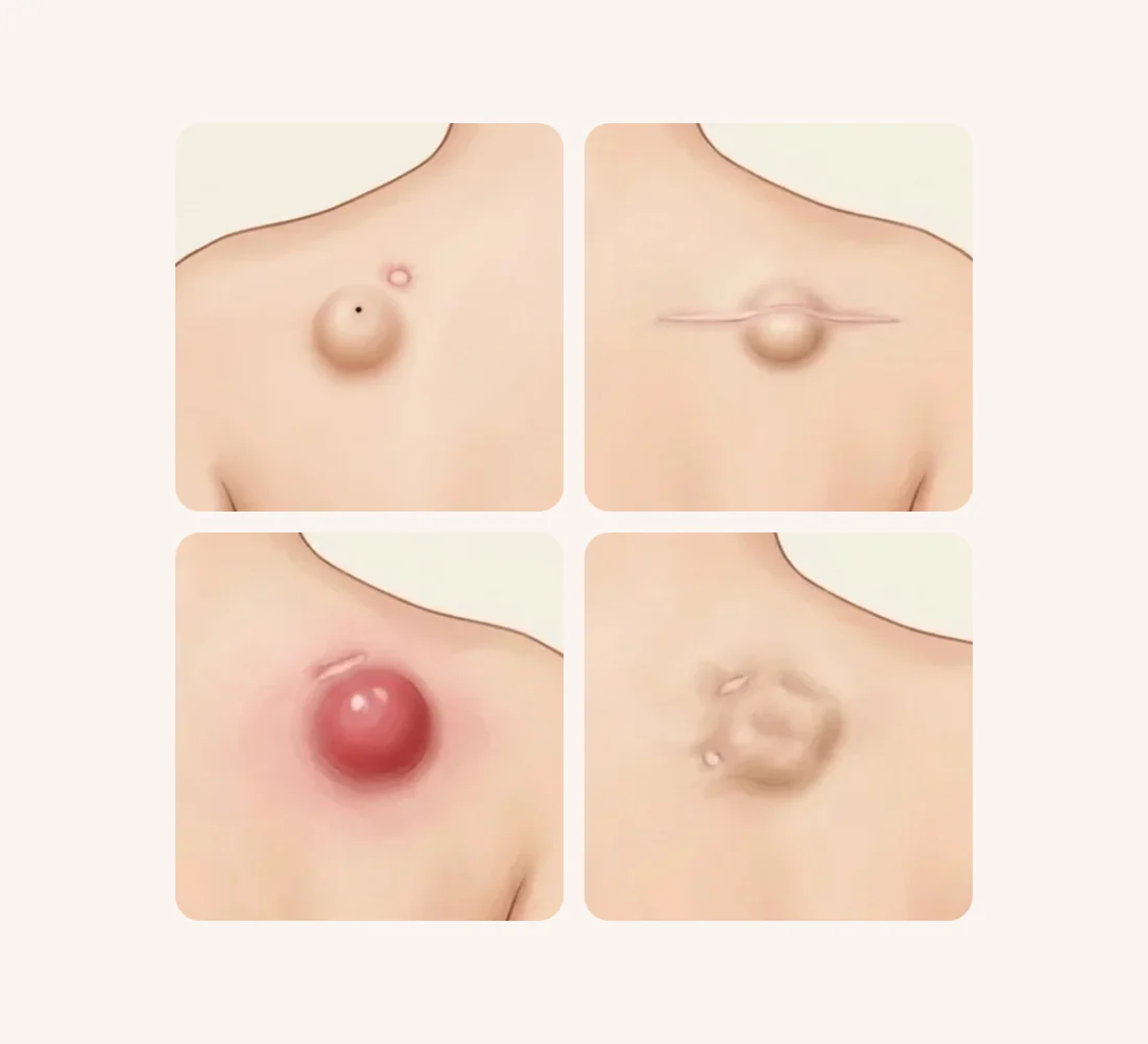
Why does a cyst come back in the same place?
To understand why a sebaceous cyst returns, it helps to know what it is made of. Most lumps people call "sebaceous cysts" are epidermoid cysts: a small sac under the skin that fills with keratin (the material skin sheds) and oily material.1
What matters is the wall of that sac. Its inner lining is made of cells much like the surface of your skin. Just as your skin keeps producing keratin and shedding it, the lining of the sac keeps producing keratin, but it has nowhere to go, so it builds up inside. That is why a cyst refills in the same spot if the wall is left behind, no matter how thoroughly the contents were emptied.
Put simply, the cause of the cyst is not what is inside it but the sac that makes what is inside it. Incision and drainage alone frequently leads to recurrence, and removing the whole sac is what prevents it.1 A systematic review (a study that gathers and compares earlier studies using a set method) likewise concluded that recurrence is related more to whether the wall was removed in full than to the size of the incision.4
A new cyst appearing somewhere else is not the same as a recurrence. A lump that appears away from where the earlier one was removed is more likely to be a new cyst. Telling us where and how earlier cysts were treated helps us tell the two apart.
Squeezing, draining, removing the sac: what is the difference?
All three make the lump smaller for a while. The difference is what happens to the sac wall, and that is exactly where recurrence and scarring part ways.
Does the sac wall stay behind?
| Method | Sac wall | Recurrence | Good to know |
|---|---|---|---|
| Squeezing | Stays in place | Refills easily | Bursting under the skin can cause inflammation |
| Incision and drainage | Usually stays | Frequent | A first step when pus has built up and it hurts |
| Removal through a small incision | Taken out | Low if the whole wall comes out | A thin wall may come out in pieces |
| Elliptical excision | Taken out whole, under direct view | Low if the whole wall comes out | Longer wound |
- Squeezing: The contents come out, but the sac stays under the skin. The lump may look smaller, yet the wall fills it with keratin again.
- Incision and drainage: This relieves pressure when a cyst is red, swollen and full of pus. It can ease the pain, but on its own it frequently leads to recurrence.1 In a systematic review, complete excision including the wall was consistently associated with fewer recurrences than incision and drainage.4 Most of the included studies were observational, though, so the review could not combine their numbers into a single estimate.4
- Excision with the sac: The sac is taken out through an incision, wall and all. One approach makes a small opening, empties the contents and then draws the wall out; another removes a small ellipse of skin together with the whole sac. In a study that randomly assigned 60 patients with uninflamed cysts to one approach or the other, recurrence did not differ meaningfully between the two.3 In plain terms, whether the wall came out completely matters more than whether the incision was small or large.
Does removing the sac mean it can never come back?
Not necessarily. Removing the whole sac is the principle for reducing recurrence, but a piece of wall is more easily left behind in these situations:
- When the wall is thin and tears: A thin wall can tear during removal and come out in several pieces. That is why surgeons are advised to inspect the wound at the end and even piece the wall back together to confirm it has all been removed.1
- When the cyst was inflamed or has burst before: Such cysts may have scar tissue around them. When scar tissue grips the wall, removing it through a small opening can be difficult.1
- When it was squeezed or drained before: The remaining wall may be collapsed and stuck to the surrounding tissue, making its edges harder to find than in an untouched cyst.
There are also numbers on how often cysts come back. In a review of cysts removed by punch incision (a small round opening) at one US dermatology practice, recurrence was 3.6% according to the clinic's records and 8.3% among patients who answered a follow-up survey.2 By site, the back (13.8%) and the ear (13.0%) showed a trend toward higher recurrence than other areas.2 In other words, most cysts did not come back, but roughly one in ten could, and the back and ears deserve a little more watching.
These figures come from one practice's past records and a survey. People who did not answer the survey are not counted, and the numbers cannot simply be applied to other methods or other sites.
When would a recurrence show up?
In the same study, 54.5% of the cysts that came back did so within the first year after surgery.2 The authors added that recurrent cysts often cause no pain, so patients tend to wait a long time before returning to a doctor.2
So for about a year after surgery, it is worth feeling around the incision line now and then. A round, pea-like lump under the line, a lump that keeps growing, or a dark dot reappearing in the center may suggest a recurrence. On the other hand, some firmness along the incision line for a few months is common while a wound heals. Rather than judging alone, take photos of the same spot from the same distance and bring them to your appointment so changes are easy to compare.
If it has come back, what comes first?
Even among recurrences, what comes first depends on the state of the lump right now.
A lump that came back: what to look at first
1It refilled after squeezing or draining
The original sac was never removed. It is taken out, wall and all, when not inflamed
Excision with the sac
2It came back under a surgical incision line
A leftover piece of wall is suspected. It is removed with the scar tissue along the earlier line
Excision with the scar
3It is red, swollen and painful again
The degree of inflammation is checked first. If pus has collected, draining it may come first
Inflammation first
4It has burst or been drained many times and feels firm and uneven
Scar tissue may be widespread. A wider excision is planned rather than a small opening
Planning the extent
- It refilled after squeezing or draining: This is less a recurrence than the original sac filling up again. The usual principle is to remove it, wall and all, at a time when it is not swollen or painful.1
- It came back under a surgical incision line: A piece of wall left from the earlier operation may be the cause. A recurrent cyst is usually removed by standard excision rather than through a small opening.1
- It is red, swollen and painful: Excision is harder while inflammation is strong, so one common approach is to wait about a week for it to settle before removal.1 On the other hand, in a study from two Korean hospitals in which 444 cysts (254 of them inflamed) were removed and closed straight away regardless of inflammation, the overall complication rate was 2.5%, with no meaningful difference between inflamed and uninflamed cysts.5 Because it looked back at past records, it does not apply to every case. Whether to remove right away or after the inflammation settles is decided by the degree of swelling and the site.
- It is firm and uneven after repeated bursting or drainage: Scar tissue may have spread widely around it. Such areas are often hard to treat through a small opening, so an excision that includes the scar tissue is planned.1
How is a recurrent cyst removed again?
Removing a recurrent cyst means dealing with two things: the leftover wall, and the scar tissue that holds it. It takes one more consideration than the first operation.
- The extent is checked by examination. The size and depth of the lump, how it relates to the old incision line, and how firm the surrounding tissue is are assessed by touch and by looking.
- After local anesthesia, the incision follows the earlier line. Following the old scar is a common way to avoid adding a second scar.
- The scar tissue and the remaining wall come out together. Pieces of wall can hide inside scar tissue, so the two are often removed as one block.
- The wound is closed in layers. Care is taken that blood or fluid does not collect in the space left behind.1
If the lump feels solid rather than cystic, is irregular in shape, or tissue other than a cyst is seen during surgery, formal excision and a tissue examination (biopsy) are recommended. Rarely, other lumps can look like a typical cyst.1
Will a second removal leave a bigger scar?
To be honest, the incision may be longer than the first time. A lump that could once be removed through a small opening may, after recurrence, need to come out together with scar tissue, which tends to widen the incision.1 If the incision follows the earlier line, however, you end up with one line rather than two.
What affects the scar:
- The size of the lump and how far the scar tissue has spread
- The site: on areas such as the back, where skin moves and stretches a lot, scars tend to widen
- Whether removal happens during inflammation or after it settles
- Whether you tend to form thick, raised scars
That is why the first operation matters most for preventing recurrence. Removing the sac, wall and all, while it is small, before it bursts and when it is not inflamed usually allows a shorter incision. If the incision line left afterward bothers you, see when to consider treatment for a surgical scar.
What matters most the first time a cyst is removed?
- Not squeezing it: Squeezing leaves the sac behind, and a rupture under the skin causes inflammation and scar tissue that make later removal harder.1
- Removing it when not inflamed: Without inflammation, the edges of the wall are clearer.
- Checking that the whole wall came out: Inspecting the removed wall and the wound is an important step in preventing recurrence.1
- Sharing the history: Telling us about earlier squeezing, drainage or surgery helps us anticipate leftover wall and scar tissue.
- Watching for about a year: More than half of recurrences were reported within the first year, so check around the incision line from time to time during that period.2
How we approach it at Bonita Clinic
At Bonita Clinic, we also see lumps that have come back after being squeezed or operated on elsewhere. Telling us where the lump is, when it appeared, whether its size has changed and whether it has ever been swollen or inflamed makes the consultation more accurate. What is possible, the method and the recovery schedule are explained at consultation.
A lump that came back: what state is it in now?
- Refilled, not inflamedRemove the sac wall and allIncision along the earlier line
- Red, swollen and painfulCheck the inflammation firstDecide: remove now or after it settles
- Firm under the scarCheck how far scar tissue extendsPlan a wider excision
Treatment options you can discuss in Seoul
Bonita Clinic opened in 1995 and has remained in the same place since, with Dr. Kwang-Seop Jung, a Brazilian board-certified plastic surgeon, seeing patients. For sebaceous cysts, our treatment page describes:
- Surgical removal under local anesthesia, with same-day discharge and stitches
- Removal of the cyst together with its sac wall to reduce the cause of recurrence
- Short incisions placed along the skin creases, including on the ears, forehead and neck
- Follow-up of the incision line at our Scar Clinic
Drainage, biopsy and the other steps described in this article are general medical information. Whether any of them is needed in your case is decided after an examination.
Why do the plan and cost differ for a recurrent cyst?
Two recurrent cysts that look alike in a photo can need quite different procedures. What has happened to the lump before, as much as its size, shapes the plan and the cost.
- Size and depth of the lump
- How far scar tissue has spread
- Earlier squeezing, drainage or surgery
- Current inflammation
- Site: back, ear, neck or face
- Stitches and follow-up visits
Final plan and quote after seeing you in person
A final estimate is given only after an in-person examination, once the size of the lump and the extent of the surrounding scar tissue are clear.
What can photos show, and what needs an in-person exam?
Photos help us see where the lump is, how the old scar looks and how things have changed. They cannot show how much scar tissue lies under the skin.
Photos can show
- Where the lump sits relative to the old scar
- Visible size, redness or a dark dot
- Squeeze or drainage marks
- Change over time (dated photos)
Needs an in-person exam
- Depth of the lump
- How firm the surrounding scar tissue is
- How freely the lump moves
- Whether inflammation is active
- Whether it is a cyst or something else
Helpful photos: the lump and the old incision line from the front and from the side, with a ruler or coin next to it for scale, in good light. Photos from when it was swollen, and the date of any earlier surgery, help too. You can send us a message with your photos and a short history of the lump.
Planning your visit and recovery
What is confirmed in advance is limited to the treatment information above: surgery under local anesthesia, same-day discharge and stitches, with stitch removal and follow-up visits explained after the procedure. How long you should stay in Seoul, and whether you need to come back for stitch removal, depend on the site and size of the lump and on how much scar tissue is removed. These are planned with you at consultation.
If the cyst is inflamed when you arrive, removal may need to wait until the inflammation settles, which typically takes about a week.1 So if the lump is currently red or painful, it is worth sending photos first.
After you get home, see a doctor promptly if the wound becomes redder, warmer, more painful or swollen, or starts to leak. Because more than half of recurrences were reported within the first year,2 dated photos of the incision line taken now and then during that year make it easier for any doctor to judge what is happening.
Real treatment cases
These cases appear on Bonita Clinic's sebaceous cyst and lipoma removal page. The ear is among the sites where recurrence was reported more often.2 Whether these were recurrent cysts cannot be told from the photos, so only the location is described. Results vary from person to person.
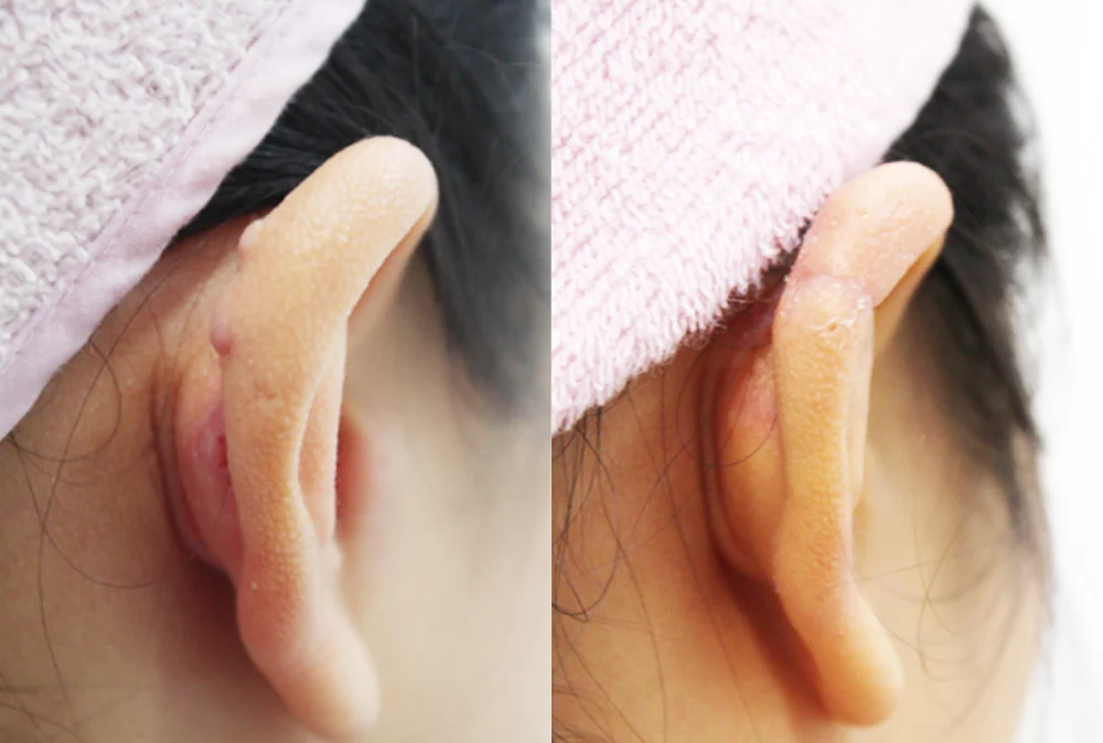
Small lumps raised along the back edge of the ear rim
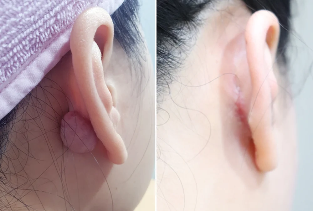
A reddish lump behind the ear
* Results vary from person to person. Side effects are possible, so please decide after a thorough consultation with a doctor.
Frequently asked questions
If it is removed again, will it definitely not come back this time?
No one can promise that exactly, but it does not mean the cycle will repeat either. If a leftover wall is the cause, removing that wall together with the surrounding scar tissue can lower the chance of it returning.4 Where scar tissue is extensive or the wall tears into thin pieces, a small fragment may still be left, so progress is followed after surgery as well.
A new lump appeared somewhere else. Is that a recurrence?
Usually not; it is more likely a new cyst. A recurrence appears where wall was left behind. People prone to cysts can develop them in other places too, such as the trunk, neck, face or behind the ears.1
Can a recurrent cyst be removed through a small opening?
Sometimes, if there is little scar tissue and the lump is small. But cysts that were inflamed or burst before often have scarring that makes removal through a small opening difficult, so standard excision is usually recommended.1 Which suits you is decided by examining it.
Can I just leave a recurrent cyst alone?
If it is small and not bothering you, watching it rather than removing it right away is sometimes an option. But if it grows or bursts, inflammation and scar tissue increase and a later incision may be longer.1 If it changes in size or starts to swell, that is a good time to seek advice.
It was squeezed a long time ago. Can it be removed now?
Yes. A time when it is not inflamed may actually be a good time to remove it. Even if it was squeezed long ago, the remaining sac is the cause, so it is removed wall and all. Telling us when and how it was squeezed helps us estimate how much it may be stuck to the surrounding tissue.
What to prepare before your consultation
- When the lump first appeared and how it has been treated: squeezing, drainage or surgery, and when
- When you first noticed it coming back, and how its size has changed since
- Any swelling or pain, and antibiotics you have taken
- Photos that show the old incision line (with a ruler or coin for scale)
- Whether you have ever formed thick, raised scars or keloids
Bonita Clinic is on the 3rd floor of 565 Gangnam-daero, Seocho-gu, Seoul, about 100 m on foot from Exit 8 of Nonhyeon Station (Line 7). You can read about the procedure on our sebaceous cyst and lipoma removal page and about scar treatment on our Scar Clinic page. Whether a lump needs removing at all, and how a first removal is done, is covered in sebaceous cysts and lipomas: why the sac matters.
References (5)
Sources that support the explanations in this article. Study findings describe general trends and do not guarantee individual results.
- Zuber TJ. Minimal excision technique for epidermoid (sebaceous) cysts. Am Fam Physician. 2002;65(7):1409-1412.Review: drainage alone frequently leads to recurrence and removing the whole sac prevents it; a thin wall may tear and come out in pieces, so the wound is inspected at the end; cysts that were inflamed or ruptured may have scarring that precludes minimal excision; recurrences are treated by standard excision; inflammation typically settles in about a week; solid tumors can masquerade as cysts
- Mehrabi D, Leonhardt JM, Brodell RT. Removal of keratinous and pilar cysts with the punch incision technique: analysis of surgical outcomes. Dermatol Surg. 2002;28(8):673-677.doi:10.1046/j.1524-4725.2002.02020.xRetrospective review with survey (abstract): recurrence after punch-incision removal was 3.6% in chart review and 8.3% among survey responders, trending higher on the back (13.8%) and ear (13.0%); 54.5% of recurrences occurred within the first year; recurrent cysts are often asymptomatic
- Lee HE, Yang CH, Chen CH, et al. Comparison of the surgical outcomes of punch incision and elliptical excision in treating epidermal inclusion cysts: a prospective, randomized study. Dermatol Surg. 2006;32(4):520-525.doi:10.1111/j.1524-4725.2006.32105.xRandomized study of 60 patients with uninflamed epidermoid cysts (abstract): no significant difference in recurrence between punch incision and elliptical excision, supporting that complete wall removal matters more than incision size
- Guru Naidu S, Can Guru Naidu N, Borgas P, Balasubramaniam D. Surgical Excision Versus Incision and Drainage for Epidermoid (Sebaceous) Cysts: A Systematic Review. Cureus. 2026;18(1):e102434.doi:10.7759/cureus.102434Systematic review of five studies (full text): complete excision including the wall was consistently associated with lower recurrence than incision and drainage, and recurrence related to incomplete excision rather than incision size; mostly observational studies, so no meta-analysis
- Gwak H, Lim ST, Kim SH. Microbiological and clinical outcomes of single-stage minimal excision of inflamed epidermal cysts without routine antibiotic treatments: A dual-center retrospective cohort study. Medicine (Baltimore). 2026;105(19):e48589.doi:10.1097/MD.0000000000048589Two-center Korean retrospective cohort of 444 cysts, 254 inflamed (abstract): immediate excision and closure regardless of inflammation had an overall complication rate of 2.5% with no difference by inflammation; most cultured bacteria were skin commensals
About this article
This article separates general medical information from Bonita Clinic's own approach. Because the right decision depends on each person's condition, it is not a substitute for a consultation.