KOKorean
 ENEnglish
 JP日本語
 CN中文(简体)
 TW中文(繁體)


Earlobe Keloid Removal in Seoul: Will a Piercing Keloid Come Back?
It can come back. Ear keloids often recur if they are only cut out, so the usual approach is to add adjuvant treatment such as injections from the time of removal and to follow the site for months to years. Adjuvant treatment does not remove the risk, which depends on the keloid's size, earlier recurrence and family history, so if you are considering treatment in Seoul, photos of the front and back of your ear are a good place to start. There is no set rule for re-piercing; it is decided after the treated site has stayed stable for a long time and the risk factors have been weighed.
Curious about your own case? Ask us online →
Why do keloids form after ear piercing?
Piercing an ear makes a small wound that runs from the front of the earlobe to the back. In most people that little channel heals without problems. In some, though, the scar tissue does not stop growing once the wound has closed, and it builds into a firm lump. That lump is a keloid. A keloid is a scar that grows beyond the edges of the original wound, so even though a piercing hole is only as wide as a needle, the lump can become many times larger than the hole.
The ear is a common place for keloids, and they usually follow an injury or an ear piercing.3 A systematic review (a review that searches for relevant studies using a set method and summarizes them) of 26 studies on ear keloids found that 15 of them named piercing as the cause. Within the ear, the earlobe was the most common site, which the authors put down to the fact that earlobes are pierced far more often than other parts of the ear.1
A hospital in Korea looked back at 40 earlobe keloids in 36 patients (the "40-keloid study" below). It included patients who had been pierced outside a medical setting, at a jewelry shop, at home or at school. All of them reported that the piercing had become swollen, inflamed or infected with pus before the keloid appeared. The lump usually began to rise 1–6 months after the inflammation settled, and it then kept growing for 1–2 years. Almost all of the patients (97%) were under 40. The authors suggested that inflammation after piercing in unhygienic conditions may be the cause.5 Because the study had no comparison group, though, it cannot prove the cause.
Because the piercing is a channel through the lobe, a keloid can form on both the front and the back of the earlobe, with the two lumps joined through that channel. One explanation is that if part of the channel is left behind during surgery, it may become a source of regrowth, although this idea has not yet been properly tested.1
Not every raised scar is a keloid. It may be a hypertrophic scar (a thickened scar that stays within the original wound). How to tell the two apart is covered in our guide to keloids versus hypertrophic scars, and keloid treatment in general in our guide to keloid and burn scar treatment. This guide focuses on piercing keloids of the ear, and of the earlobe in particular: what makes them come back after removal, and what lowers that risk.
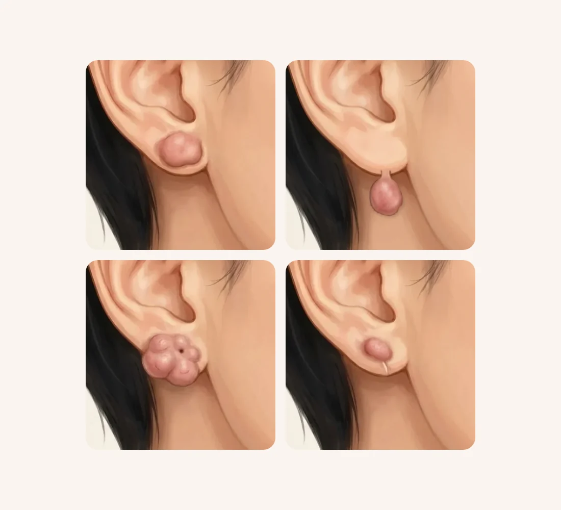
What shapes can an earlobe keloid take?
Shape matters first because it changes how a keloid is removed. A university hospital in Korea classified 1,027 earlobe keloids it had treated over 10 years by their outward appearance. It sorted them into five types, listed here from most to least common:6
- Sessile, single nodule: one lump attached to the lobe over a broad base
- Pedunculated: a lump hanging from the lobe by a narrow stalk
- Sessile, multinodular: several lumps merged over a broad base
- Buried
- Mixed
Earlobe keloids: which shape is yours?
1One lump broadly attached at the piercing
The most common shape. Its size and front-to-back extent guide whether injection or excision is considered
Sessile, single nodule
2A lump hanging from the lobe on a narrow stalk
The extent of the stalk is checked, and recurrence control is planned together with any excision
Pedunculated
3Several bumps merged into one lumpy mass
How far it spreads and whether it reaches the lobe edge decide how it is removed
Sessile, multinodular
4A lump back on the line of an earlier removal
Treated as a recurrence: settled first with injections, with any repeat excision weighed carefully
Recurrence
The researchers chose a different operation for each shape: removing only the core of the lump, cutting out the whole lump, or a combination of the two. They reported closing every case directly, without skin grafts and without sacrificing the surrounding tissue.6 The 40-keloid study used a similar rule. If the lump stayed around the piercing site, only its core was removed; if it had spread to the edge of the earlobe, it was cut out.5
In short, the shape is a clue to how the keloid should be taken out. So the first step is to confirm that it is a keloid and look at its shape, and then to decide whether to settle it with injections or to plan excision together with recurrence control. Whatever the method, a keloid can grow back where it was removed, which is why care after removal is planned from the start.
Why does a keloid come back if it is only cut out?
Cutting out a keloid is itself a new wound. When a new wound is made in someone whose skin tends to form keloids, at a site where a keloid has already formed, the scar tissue can overgrow again while that wound heals. In older reports summarized in the introduction of a randomized trial (a study that divides patients into groups at random), keloids removed by excision alone came back in 45% or more of cases, while adding steroid injections after surgery brought recurrence below 50%.4 By those reports, cutting a keloid out without anything else left close to half or more at risk of regrowth. A recent meta-analysis (a study that pools the results of several studies and recalculates them) limited to earlobe keloids, however, found that the estimated recurrence after excision alone could be similar to that of combination treatments, so this figure varies widely between studies.2
Inflammation often lingers in earlobe keloids, too. In the 40-keloid study, when the removed tissue was examined under a microscope, 70% of the 40 keloids showed inflammatory cells gathered around blood vessels both inside the keloid and in the tissue around it. Three of the four keloids that came back appeared to be linked to inflammation that continued after surgery.5
For the same reason, our treatment page explains that a keloid can come back larger if it is excised carelessly, so we think about controlling it before cutting it. When removal is needed, reducing the tension on the stitches and continuing recurrence management afterwards are treated as one course of care.
What adjuvant treatments are used after removal?
An adjuvant treatment is an added treatment that holds back overgrowth of scar tissue while the excision site heals. In the review of 26 studies, the adjuvants used most often were dressings and products applied to the skin, including pressure and silicone (37.9%), steroid injections (22.2%), radiotherapy (17.6%) and carbon dioxide laser (10.5%).1 The illustration below shows how recurrence control follows an excision.
Steroid injections: the timing matters
A steroid injection calms the inflammation inside a keloid and reduces the production of scar fibers. It is regarded as the first-line treatment for an established keloid, and for ear keloids, injection on its own or injection combined with excision are the most common approaches.4
In a meta-analysis of 25 studies on ear keloids, recurrence after excision followed by steroid injection was 15.4%.3 That works out to roughly one person in six or seven.
The randomized trial mentioned above was a small one that compared the timing of the injection. Of 34 patients with ear keloids, one group received an injection into the wound straight after the keloid was removed and stitched, and the other received it one week later. Within 6 months, 22.2% of the immediate group had a raised regrowth, compared with 56.3% of the one-week group. No complications such as infection or thinning of the skin were seen.4 The trial was small, however, and followed patients for only 6 months. The authors also noted that they used strict criteria to catch recurrence early, which made the recurrence rates high.4
More injections are not automatically better. Steroid injections can thin the skin, leave pale patches or make small blood vessels visible, so the dose and spacing are adjusted.4
Pressure earrings: consistency is key
Pressure therapy uses a magnetic or clip-style pressure earring to squeeze the treated earlobe from front and back. It is thought to work by reducing blood flow to the scar, which slows down the cells that make scar fibers.1 In the 40-keloid study, patients were asked to wear magnetic pressure earrings from 2 weeks after surgery for 6 months and to use a scar ointment, and 10% (4 keloids) came back.5
Across studies that used pressure as an adjuvant, though, recurrence ranged widely, from 0% to 29.5%. Wearing a device for long periods can make the ear sore and can cause pressure sores. In one study of keloids on both ears, where patients alternated the pressure clips between ears, recurrence was higher.1 In practice, the benefit depends heavily on how long and how consistently the pressure is kept up. Pressure therapy is not listed on Bonita Clinic's treatment page, so it is described here as general medical information.
Radiotherapy and other options
Recurrence after excision followed by radiotherapy was 14.0%, with no clear difference from steroid injection (15.4%).3 In the review of 26 studies, recurrence with radiotherapy as an adjuvant mostly stayed at or below 8.7%, although one trial reported 56.7%.1 Reported side effects were mainly redness of the treated skin, and with appropriate technique the risk of cancer was reported to be very low.1 Even so, it means using radiation for a benign condition, so it is a treatment decided with care. The earlobe keloid meta-analysis mentioned above (85 studies) found that the estimated recurrence rates of six approaches, excision alone included, looked similar to one another. Within radiotherapy, electron-beam treatment showed possible advantages over X-ray treatment, which partly explained why results differed between studies. When the data from individual patients were pooled and analyzed again, however, both excision with steroids and excision with radiotherapy gave longer recurrence-free periods than excision alone. Rates of adverse events were similar across the treatments.2 Radiotherapy is not on Bonita Clinic's treatment page, so please ask at consultation whether it might be relevant to you.
Another option is to mix 5-FU (a drug that slows cell growth) with the steroid injection. In one randomized trial, patients who received this injection after excision had fewer recurrences than those who received radiotherapy; wound separation and redness were reported in a few patients in that trial.1 Using a carbon dioxide laser followed by a vascular laser instead of surgery has been reported only in case series, one of which was later retracted, so the evidence is limited.1
Adjuvant treatments after removal: what studies show
| Adjuvant | What studies found | Keep in mind |
|---|---|---|
| Steroid injection | 15.4% recurrence after excision (meta-analysis); fewer if given at closure than at 1 week | Thinning, pale patches or visible small vessels can occur |
| Pressure earrings | 0–29.5% recurrence across studies, a wide range | Worn for months; pain and pressure sores possible |
| Radiotherapy | 14.0% recurrence after excision (meta-analysis), no clear difference from injection | Not on our treatment page (general information); mainly redness reported |
| 5-FU added to injection | Fewer recurrences than radiotherapy in one randomized trial | General information; wound separation and redness reported in that trial |
How should you read recurrence rates?
The figures in studies cannot simply be turned into your personal odds. The chart below brings together the numbers mentioned in this guide.
In the review of 26 studies, the average recurrence rate for ear keloids was 20.3%, and 19.4% when only studies that combined surgery with an adjuvant were counted.1 Put simply, even with adjuvant treatment, on average roughly one person in five may see the keloid return. The rates varied a great deal between studies, however, because their size, follow-up and treatments all differed. Follow-up ranged from 6 months to 6 years, with an average of 25.8 months. Most studies were of low to moderate quality, and only one included more than 100 patients.1
What matters more than any single number is which risk factors your own keloid has. In studies of piercing keloids, outcomes were shaped mainly by the size of the keloid, whether it had recurred before, and whether keloids ran in the family.1 The authors of the earlobe keloid meta-analysis likewise concluded that each patient's overall condition should be weighed, and that more studies with long enough follow-up are needed.2
When does recurrence show, and what are the warning signs?
Recurrence often shows up months after surgery rather than straight away. In the injection-timing trial, new regrowths kept appearing between the second and fifth months after surgery.4 Just as the original keloid took 1–2 years to grow,5 regrowth can take time as well. Across ear keloid studies, the average follow-up was a little over two years (25.8 months).1
These changes may be a sign of recurrence:
- The treated site becomes firm again, or starts to rise
- The site itches, or hurts when pressed
- Redness spreads around the stitch line, and the red rim widens
The 40-keloid study also watched for firmness or redness at the treated site as a sign that a keloid might be returning.5 In the randomized trial, keloids that grew back were injected at monthly intervals.4 Noticing regrowth early means it can be dealt with straight away, for example with injections, so dated photos taken from the same angle and in the same light are useful for comparison.
Pregnancy is also worth considering. In the 40-keloid study, one of the four keloids that came back did so with each of the patient's pregnancies, which the authors attributed to hormonal influence.5 That is a single case, so it cannot be said to happen in every pregnancy, but if you are planning a pregnancy it is worth discussing the timing of treatment in advance.
Can I pierce my ears again after a keloid?
We could not find a guideline or a large study that sets out whether, or when, re-piercing is advisable after a keloid has been treated. What follows is therefore a set of points to think through in light of the risk factors that studies have identified. Remember first that re-piercing means making a new wound in the same skin.
- Has the treated site settled for long enough? Recurrence often appears months later,4 and the average follow-up in studies was a little over two years.1 If the site is still red, itchy or firm, treatment and observation come before any new piercing.
- Do several risk factors overlap? Keloid size, earlier recurrence and family history are named as the main factors shaping outcomes in piercing keloids.1 If the keloid has recurred more than once, or keloids run in your family, more caution is needed, and not re-piercing is also an option worth considering.
- Can inflammation be prevented? In the 40-keloid study, every patient had inflammation at the piercing before the keloid appeared.5 If you do re-pierce, choose a clean, hygienic setting, and if the site swells or produces pus, get it treated promptly rather than waiting.
- Where should a new piercing go? We could not find studies on this. Making a new hole in the scar left after a keloid was removed, however, adds a wound to a place where regrowth can already happen, so please decide the site together with your doctor at an examination.
Before re-piercing, how is the treated site?
- Still red, itchy or firmTreatment and observation firstCheck whether it is a sign of recurrence
- Unchanged for a long timeWeigh the risk factors togetherPast recurrence, family history, earlier size
- Recurred more than once, or family historyBe more cautiousNot re-piercing is also an option
If a new piercing starts to become firm, itchy or red, take the earring out and have it examined early. It is also a good idea to tell whoever does the piercing that you have been treated for a keloid before.
How we approach it at Bonita Clinic
How we proceed at Bonita Clinic
- 1Check the scar typeKeloid or hypertrophic scar, size and extent
- 2Injection treatmentSettles red, firm, raised keloids
- 3Low-tension excisionIf excision is needed, recurrence management follows
- 4LaserWhen redness, pigment or texture remains
- 5Progress checksSpaced visits, adjusting the method
Injections are repeated at intervals to manage recurrence
At Bonita Clinic, keloid treatment is given as a procedure, with excision if needed. It is done under local anesthesia, and you go home the same day; stitches are used only if excision is done. How many surgical and procedural restoration sessions are needed varies from scar to scar and is decided as healing is reviewed, and recovery time and follow-up visits also depend on the extent of treatment and your condition. Bleeding, infection, inflammation and other side effects can occur after surgery or a procedure.
Treatment options you can discuss in Seoul
Bonita Clinic opened in 1995 and has stayed at the same location since, where Dr. Kwang-Seop Jung, a plastic surgeon board-certified in Brazil, sees patients. The clinic is on the 3rd floor of 565 Gangnam-daero, Seocho-gu, about a 100 m walk from Exit 8 of Nonhyeon Station on Line 7. For an ear keloid, the options discussed in consultation are the ones on our treatment page:
- Injection treatment for red, firm, raised keloids, repeated at intervals
- Excision with low-tension suturing, followed by recurrence management
- Combined lasers for remaining redness, pigmentation and texture
Which of these fits, and in what order, depends on the size, shape and extent of your keloid and on how it has behaved so far, and that is confirmed by examination.
Why the plan and cost differ from one ear keloid to another
A small single lump on one earlobe and large keloids on the front and back of both ears are very different situations and call for different plans. The size and number of keloids, whether they sit on one side or both sides of the lobe, the methods combined and the number of sessions all shape the plan, and with it the cost.
- Size, number and shape
- Front, back or both sides
- One ear or both
- Injection, excision or both
- Number of sessions and spacing
- Earlier treatment and recurrences
Final plan and quote after an in-person exam
A final estimate is given only after an in-person examination, once the extent of the keloid, the methods and the likely number of sessions are clear. Photos help narrow down the options, but the scope is set in person.
What photos can show, and what needs an in-person exam
Photos are a useful first step for an ear keloid, because where it sits, how large it is and what shape it has are often visible. How firm and deep it is, and whether lumps on the front and back are connected through the piercing channel, cannot be judged from a screen.
Photos can show
- Location on the front or back
- Size (with a ruler) and color
- Shape: broad, hanging or lumpy
- Change over time (dated photos)
Needs an in-person exam
- Firmness and depth by touch
- Whether front and back lumps connect
- Itch, pain and tenderness
- Scars from earlier surgery
- Signs of a different growth
Helpful photos: the front and the back of the ear in daylight without filters, a close-up of the keloid with a ruler or coin beside it for scale, and any older photos showing when it first appeared. If it has been treated before, a photo of the site right after that treatment helps too. You can send us a message with your photos, when and where the ear was pierced, and the treatments you have had so far.
Planning your visit and follow-up after you return home
What is confirmed in advance is limited to the treatment information above: a procedure with excision if needed, local anesthesia, same-day discharge, and stitches only if excised. Injections are repeated at intervals, so how many times you need to come to Seoul, and how long to stay, are planned with you at consultation, according to the size, shape and extent of your keloid.
Ear keloids call for long follow-up, often well beyond the treatment visits themselves; in studies, recurrence often appeared months after surgery,4 and the average follow-up was a little over two years (25.8 months).1 Before you leave Seoul, ask which signs to watch for, such as new firmness, itch or a widening red rim, and how follow-up treatment would be arranged if the keloid starts to come back after you return home. Keep taking dated photos from the same angle so that changes can be compared with your consultation photos. If the treated area becomes painful, red and hot, or starts to discharge, see a local doctor promptly.
Real treatment cases
These ear cases appear on Bonita Clinic's burn and keloid treatment page. Photos alone cannot show whether a piercing was the cause, or whether each lesion was a keloid or a hypertrophic scar, so only what is visible is described. Results vary from person to person.
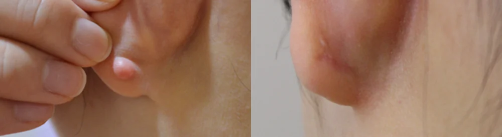
A small, rounded raised lump on the lower part of the earlobe
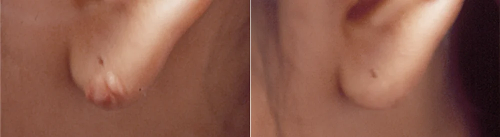
Small raised lumps along the lower edge of the earlobe
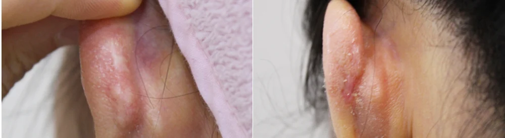
A broad, reddish-purple raised lump on the back of the ear
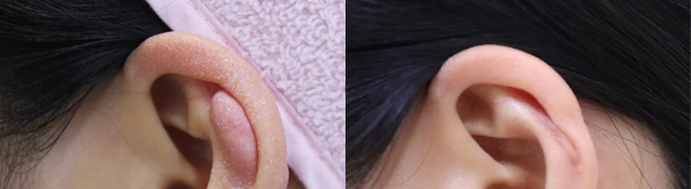
An elongated raised lump on the inner part of the upper ear
* Results vary from person to person. Side effects are possible, so please decide after a thorough consultation with a doctor.
Frequently asked questions
Can an ear keloid be treated with injections alone?
Small keloids sometimes shrink with injections, and steroid injection is regarded as the first-line treatment for an established keloid.4 Whether injections alone will be enough, or whether excision with recurrence control is needed, depends on the size, the shape and how the keloid responds to injections. At Bonita Clinic, red, firm, raised keloids are injected at intervals while progress is followed.
Do I have to wear a pressure earring after surgery?
Pressure is one of several adjuvant treatments used to lower recurrence. In the Korean study, where patients wore magnetic pressure earrings for 6 months, 10% of keloids came back,5 but results varied widely between studies, and soreness or pressure sores can occur.1 Pressure therapy is not listed on Bonita Clinic's treatment page, so whether it is needed is discussed at consultation alongside other ways of managing recurrence.
If the keloid comes back, will I need surgery again?
When a regrowth is still small, settling it with injections is usually considered first. In the randomized trial, keloids that grew back were injected at monthly intervals.4 Because earlier recurrence is itself one of the factors that shape outcomes,1 any repeat excision is planned together with adjuvant treatment and long follow-up from the start. Cutting out the same site again and again without added treatment is avoided.
Can pregnancy make a keloid grow back?
There are reports that it can. In the Korean study, one of the four keloids that came back did so with each of the patient's pregnancies, which the authors put down to hormonal influence.5 It is a single case and cannot be generalized, but if you are planning a pregnancy, please discuss the timing of treatment and follow-up in advance.
Is a keloid on an upper-ear (cartilage) piercing the same?
Piercing keloids also form on the upper rim of the ear (the helix). In the injection-timing trial, 53% of keloids were on the helix and 44% on the earlobe, and 94% followed a piercing.4 Many studies mix earlobe and helix keloids, so their results cannot easily be compared by site.1 The principles of excision, adjuvant treatment and long follow-up are the same, but the way a keloid is removed and closed depends on its site and shape.
Before your consultation
- When and where your ear was pierced, and whether it became swollen or infected afterwards
- When the lump first appeared and how fast it has grown, and whether it is on the front, the back or both
- Treatments so far: injections (how many and when the last one was), excision, pressure earrings, laser, radiotherapy
- If it has come back before: how many times, and how long after surgery
- Whether anyone in your family has keloids, and whether you are planning a pregnancy
- Photos of the front and back of the ear in good light, plus older photos from when it first appeared
You can see the treatment process on our burn and keloid treatment page. For settled burn scars, see our burn scar page. If you are not sure whether a raised scar is a keloid, see our guide to keloids versus hypertrophic scars, and for the evidence and limits of injections, lasers and excision, our guide to keloid and burn scar treatment.
References (6)
Sources that support the explanations in this article. Study findings describe general trends and do not guarantee individual results.
- Haq H, Davies L, Dheansa B. Understanding and Comparing Current Practice for Ear Keloid Scars: A Narrative Systematic Review. J Plast Reconstr Surg. 2026;5(4):326-336.doi:10.53045/jprs.2025-0067Systematic review of 26 ear keloid studies (narrative synthesis): piercing named as the cause in 15; earlobe most common; average recurrence 20.3% (19.4% with surgery plus adjuvant); mean follow-up 25.8 months; pressure 0–29.5%, with soreness and sores; radiotherapy mostly 8.7% or less (one trial 56.7%), mainly redness, very low reported cancer risk; 5-FU combination beat radiotherapy in one trial (wound separation, redness reported); one of two CO2-plus-vascular-laser case series later retracted; outcomes shaped by size, earlier recurrence and family history; low to moderate quality
- Huang Y, Ren S, Yang Q. Efficacy and Safety of Excision Combination Therapies for Earlobe Keloids: A Systematic Review and Meta-analysis. Aesthetic Plast Surg. 2024;48(15):2757-2770.doi:10.1007/s00266-024-04092-0Meta-analysis of excision-based combination treatments for earlobe keloids (85 articles, 23 treatments): estimated recurrence of several approaches looked similar; in individual patient data, excision with steroids and excision with radiotherapy gave longer recurrence-free survival than excision alone; adverse event rates were similar; studies with sufficient follow-up are needed
- Shin JY, Lee JW, Roh SG, et al. A Comparison of the Effectiveness of Triamcinolone and Radiation Therapy for Ear Keloids after Surgical Excision: A Systematic Review and Meta-Analysis. Plast Reconstr Surg. 2016;137(6):1718-1725.doi:10.1097/PRS.0000000000002165Meta-analysis of adjuvant treatment after ear keloid excision (25 studies): the ear is a common keloid site after trauma or piercing; recurrence 15.4% with triamcinolone and 14.0% with radiotherapy, with no significant difference; larger randomized studies needed
- Burusapat C, Wanichjaroen N, Wongprakob N, et al. The Effectiveness of Immediate Triamcinolone Acetonide Injection after Auricular Keloid Surgery: A Prospective Randomized Controlled Trial. Plast Reconstr Surg Glob Open. 2021;9(8):e3729.doi:10.1097/GOX.0000000000003729Randomized trial of 34 ear keloid patients (94% after piercing; helix 53%, earlobe 44%): recurrence 22.2% with injection at closure versus 56.25% at one week (6 months, strict criteria), no complications seen, limited by small size and short follow-up; its introduction and discussion summarize 45% or more recurrence after excision alone and below 50% with postoperative injection, injection as first-line for established keloids, and local and systemic side effects of injections
- Kim HD, Chu SC, Hwang SM, et al. Clinical Analysis of Lobular Keloid after Ear Piercing. Arch Craniofac Surg. 2016;17(1):5-8.doi:10.7181/acfs.2016.17.1.5Retrospective review of 40 earlobe keloids (36 patients) at one Korean hospital: patients pierced outside medical settings (a selection criterion, no comparison group; the authors suspected inflammation after unhygienic piercing), inflammation or pus before the keloid, onset 1–6 months after inflammation settled and growth for 1–2 years, 97% under 40, core removal if confined to the piercing site and excision if it reached the lobe edge, magnetic pressure earrings from 2 weeks for 6 months, recurrence 10% (one recurring with each pregnancy), perivascular inflammation in 70%
- Park TH, Seo SW, Kim JK, Chang CH. Earlobe keloids: classification according to gross morphology determines proper surgical approach. Dermatol Surg. 2012;38(3):406-412.doi:10.1111/j.1524-4725.2011.02199.xRetrospective classification of 1,027 earlobe keloids at a Korean university hospital: sessile single nodule, pedunculated, sessile multinodular, buried and mixed types in that order of frequency; core extirpation, standard or wider excision and combinations chosen by shape, all closed directly without skin grafts
About this article
This article separates general medical information from Bonita Clinic's own approach. Because the right decision depends on each person's condition, it is not a substitute for a consultation.