KOKorean
 ENEnglish
 JP日本語
 CN中文(简体)
 TW中文(繁體)


Old Silicone and Paraffin Injection Removal in Seoul: Is It Needed When You Have No Symptoms?
Having no symptoms does not mean the material must come out right away, but "nothing has happened" is not the same as "safe". Silicone and paraffin do not dissolve in the body, so years later they can show up as a firm lump (granuloma), as material that has moved, or as late inflammation. The first step is an ultrasound to see where the material is, how much there is and how it is spread; the decision to watch or remove is then made from those findings together with the site and any change over time. If the substance is unknown, or anything is growing or hardening even slightly, it is worth having it checked without delay. If you are planning a visit to Seoul, you can send photos and what you remember about the injections before you come.
Curious about your own case? Ask us online →

How are silicone and paraffin injections different from today's fillers?
Liquid silicone and paraffin injections were procedures in which liquid silicone (silicone oil) or paraffin (a mineral oil) was injected straight under the skin to add volume. Today's most common filler, hyaluronic acid, is classed as a filler the body usually breaks down within about a year. Liquid silicone, by contrast, is classed as a filler that does not break down and stays in the tissue.1 Put simply, even if your injection was given thirty years ago, the material should be assumed to still be under your skin.
Paraffin is described as perhaps the first substance ever used as a facial filler. It caused severe granulomatous reactions and what is called a "paraffinoma".1 A granuloma is a firm lump of chronic inflammation that forms when the body walls off material it cannot dissolve. Pure silicone oil was long thought to provoke little reaction in the body. Its use as a filler became controversial, however, because of the high rate of complications reported.1
A bigger problem is that many of these injections were given without records, using substances that were not approved medical products. A study from a hospital in Korea looked at 64 people treated for foreign body granulomas of the face. The largest group, 27 people (42.2%), had been injected with an unknown, illegally administered substance.2 In other words, about four in ten did not know what was in their face when they came for help.
This guide is about the situation where you have no noticeable symptoms right now: what to check, and how the decision to watch or remove is made. If there is already swelling or a lump, our area-specific guides go into more detail: the forehead, chin and cheek filler removal guide for the face, and the guides on buttock and leg injections and breast injections for the body.
If nothing has happened for decades, can problems still appear?
The body recognizes material it cannot dissolve as "not mine". Immune cells gather around it and wrap it up, as if sealing it in a thin wall. Many people live with this quietly for a long time. But if immune cells keep gathering and the chronic inflammation continues, a firm lump you can feel, a foreign body granuloma, can form.3
A review of imaging for fillers describes these granulomas clearly. They are a chronic inflammatory reaction, not an allergy. They appear months to years after the injection and grow very slowly. There may be several, and they can come back. Under the microscope, they send finger-like extensions into the surrounding tissue.1 That last feature means their edges are unclear, which is why they are hard to remove cleanly with surgery.2
A recent study of liquid silicone granulomas makes the same points. These granulomas often appear years after the injection. And because silicone breaks up into tiny droplets that seep into nearby tissue, the authors describe removing it all surgically as nearly impossible.3
Why some people stay quiet for life while others develop problems years later is still not well understood. Factors that researchers mention include the type and purity of the substance, the amount injected, the number of injections, unknown substances, infection, and differences in how each person's body reacts.2 So "it has been fine so far" is not, on its own, a reliable forecast.
What changes can appear over time?
The review lists the following late complications.1
- A firm lump (foreign body granuloma): a lump you can feel, or an uneven contour
- Late infection: infection itself is rare, but when it occurs it can appear months to years later as swelling with pus
- Migration: the material is found outside the place where it was injected
- Skin changes: lasting discoloration, scarring, tissue death (necrosis) and wounds that do not heal (ulcers)
Material can travel further than people expect. A Korean radiology team reported mammograms of nine women who had had foreign material injected into the face and neck to smooth wrinkles. Several small white spots, each under 5 mm, appeared in the upper inner part of the breast. They were foreign body granulomas that had moved down from the face and neck.4 Findings in unexpected places can be mistaken for something else, which is one reason to tell doctors about old injections.
Rarely, the whole body is affected. A systematic review (a study that gathers all relevant research by a set method) looked at hypercalcemia, meaning too much calcium in the blood, after cosmetic injections. It found only 23 patients across 20 published articles. Silicone was the most common substance (43.5%), and the buttocks (69.6%) and breasts (39.1%) were the most common sites. The high calcium appeared on average about eight years (7.96 years) after the first procedure. Kidney failure was the most frequent complication (82.4%), and some patients died.6 This is very uncommon. But if a blood test has ever shown high calcium or abnormal kidney results, your old injection history matters and should be shared.
What people notice on the outside usually looks like one of the four situations below. The drawings show the same face with only that difference.
Which of these is closest to you?
1Nothing shows on the outside
Ultrasound records its location, amount and shape; whether to remove is discussed by substance and site
Check and record
2You can feel a firm lump
It may be a granuloma; its size, depth and edges guide the removal plan
Granuloma
3A lump has appeared below where it was injected
The material may have moved; the new and the original site are both checked
Migration
4It turns red, swollen or warm
Treat the inflammation first, then map the material once it settles
Inflammation
If it doesn't hurt, isn't it fine?
Not necessarily. There is evidence that problems from injected material often first show up as a lump that does not hurt. It comes from a study of people seen after paraffin injection into the breast, which combined 60 patients from one hospital with 31 published reports, 232 people in total. The most common first symptom was a lump without pain (65.5%), followed by a painful lump (14.7%) and an ulcer (7.3%).5 Put another way, two in three people came in because of a lump that did not hurt.
Breast paraffinoma: why people first sought care
Treatment in the same study varied widely. More than half of the reported cases (53.4%) went on to have the breast removed (mastectomy). Yet one in four (25%) needed no further treatment. The authors stressed that any breast cancer present at the same time must not be missed.5 These figures include older cases, so they do not reflect how treatment is done today. Two points are still clear. No pain does not mean no problem, and not everyone needs major surgery.
With no symptoms, what should be checked first?
The starting point is an accurate picture of what is there now. Looking and feeling only reveal lumps close to the skin. Material that sits deep or has spread widely is easy to miss. Each imaging test sees some things well and others poorly.1
- Ultrasound: a scan that looks inside without any incision. It is safe, available and done on the spot, and it is used to find material, granulomas and abscesses in the fat just under the skin. Results do depend on the skill of the person scanning.
- Ultrasound's limit: silicone shows up as a bright "snowstorm" that blurs and hides the tissue beneath it. In the Korean mammography study above, strong shadowing also hid the structures under the skin of the lower neck.4
- MRI: better for wide areas, deep tissue and material that has moved somewhere else. Silicone has a distinctive look on MRI, and a sequence exists that shows silicone alone.
- CT: granulomas caused by silicone or paraffin can show rounded, eggshell-like calcification (calcium deposits that harden like stone).
Tell doctors about old injections whenever you have other scans, too. On PET-CT, a whole-body scan, most fillers normally light up strongly. The radiologist needs to know so this is not mistaken for disease. Fillers can also hide a real problem.1 If the material is in the breast, take particular care that breast screening does not miss a cancer that may be present at the same time.5
At Bonita Clinic, before deciding on removal, we first use ultrasound to check where the remaining material is and the condition of the tissue around it. You can consult with us even if you do not know what was injected. Simply tell us as much as you remember about when, where and what kind of procedure you had.
Watch or remove: what decides it?
There is no shared standard for when to remove material that causes no symptoms. Even for treating liquid silicone granulomas, researchers note that no consensus exists yet.3 So several things are weighed together:
- What was injected? The less is known about the substance, the harder it is to predict how it will behave.
- How much, and how is it spread? Material gathered in one place gives a fairly clear removal area. Material spread widely has blurred edges and is hard to remove cleanly.23
- Is it changing? If it grows, hardens, moves or turns red, do not wait.
- Where is it? Thin skin, areas that move a lot with expression, and the breast, which needs regular screening, each raise their own points.
- How is your general health? If you have been told your calcium or kidney results were abnormal, a medical evaluation may be needed as well.6
What is the material under your skin doing now?
- Small, in one place, unchangedWhether to remove: discussed by substance and siteIf removed, the area is fairly clear
- Spread widely, unchangedHard to remove it all, so weigh gains and risksProblem areas first, in stages
- Growing, hardening or movingDo not wait: map it, then consider removal
- Red, swollen, painful or feverishInflammation is treated first; contact us promptly
For timing once symptoms have started, there is some evidence. In the Korean study of 64 patients, the earlier surgery was done after the first visit, the shorter the treatment period and the higher the satisfaction.2 That study, however, analyzed people who already had symptoms. It cannot simply be used as proof that removing material early is better for people without symptoms.
At Bonita Clinic, we recommend removal for substances of unknown composition, because they are more likely to cause inflammation or hardening. How far and when to remove, though, is decided together with you after looking at the extent seen on ultrasound, the condition of the tissue, and the shape that will be left afterwards.
If you decide to remove it, how much can come out?
Removing every last trace is not a realistic goal. As described above, granulomas reach into surrounding tissue,1 and silicone seeps in as tiny droplets, so the edges are blurred.23
Material left behind can cause trouble again. An Indonesian study followed 35 people with liquid silicone granulomas of the chin for an average of about two and a half years (137.9 weeks). Of those who had surgery alone to cut out the granuloma, 8 of 18 (44.4%) saw it come back. Of those who also received steroid injections after surgery, 1 of 17 (5.9%) did.3 In plain terms, with surgery alone it returned in more than four in ten people. Still, it is a single small study, and the groups were formed by whether patients agreed to the injections rather than at random, so it should not be over-generalized. It does show how much care and follow-up after removal matter.
After removal, the following can happen:
- Hollowing: the area can sink by as much volume as the material used to take up. That is why the need for restoration is considered when removal is planned.
- Scars: a scar can remain where the incision was made.
- Uneven contour or recurrence: the shape can be uneven after surgery, or swelling can keep coming back because of material left behind. In the Korean study of 64 patients, those who were dissatisfied reported deformity and scarring from surgery, and in some, symptoms returned from time to time.2
- Work near nerves and vessels: on the face, if the material lies deep or has spread widely, surgery reaches areas close to the nerves that move facial expression and to blood vessels, so the extent of removal is set with extra care.
- Risks of surgery itself: bleeding, infection, inflammation and other complications can occur.
At Bonita Clinic, foreign material removal is done as surgery. For the face, we use sedation combined with local anesthesia, and you go home the same day. For the buttocks and legs, sedation or general anesthesia is used, and an overnight stay may be needed depending on the extent. For the breast, general anesthesia or sedation is used, with same-day discharge or one night's stay. There are stitches, and the length of recovery and number of follow-up visits depend on the extent of treatment and on each person. For ways to address hollowing or sagging after removal, see our guide to lifting after filler and foreign material removal.
If you decide to watch it, what should you keep track of?
If you decide not to remove it now, aim for "watching with knowledge" rather than "doing nothing".
- Keep a record: store the ultrasound report, front and side photos, and the date and site of the injections together. They become the baseline for spotting change later.
- How often to check: there is no evidence for a fixed interval, so it is agreed with your doctor according to the amount and location of the material.
- Signs to act on promptly: a lump that grows or hardens; redness, swelling, warmth or pain; skin that thins or changes color; a wound that will not heal, or pus.1 Do not wait for your next scheduled check if these appear.
- Tell others before tests or treatments: before a mammogram, MRI or PET-CT, and before any other procedure on the same area, mention the old injections first. Repeated injections in the same place are also mentioned as one factor that can stir up a granuloma reaction.2
- Whole-body signs: if a blood test shows abnormal calcium or kidney results, tell the doctor in charge about your old injections.6
How we approach it at Bonita Clinic
The order we follow at Bonita Clinic
- 1Your historyWhen, where and what was injected (unknown is fine)
- 2UltrasoundLocation, extent and tissue condition
- 3Plan extent and shapeHow much to remove, whether restoration is needed
- 4RemovalWithin the mapped area, limiting tissue damage
- 5Restore and follow upYour own fat if needed; any remainder is watched
Since opening in 1995, Bonita Clinic has been at the same location, where Dr. Kwang-Seop Jung sees patients. The actual extent and method are decided after an in-person examination.
Treatment options you can discuss in Seoul
Dr. Kwang-Seop Jung holds Brazilian board certification in plastic surgery and has 31 years in plastic surgery and scar restoration. For old silicone, paraffin or unknown injections, what you can discuss with us at consultation is the approach described on our treatment pages:
- Ultrasound to locate the remaining material and check the surrounding tissue
- Surgical removal within the mapped area, with attention to the shape left afterwards
- Restoration of hollowed areas with your own fat when needed, timed by the tissue's condition
- Continued care of any remaining scar at our scar clinic
Details by area are on our pages for forehead, chin and cheek revision, buttock and leg revision and breast revision after injections. Which steps you need is decided only after an examination.
Why do the plan and cost differ from person to person?
Two people who both say "I had silicone injected years ago" can need very different treatment. What shapes the plan, and therefore the cost, is what was injected, how far it has spread and what the area needs afterwards.
- What was injected, and how long ago
- How far it has spread, and how deep
- Where it is: face, breast or buttocks and legs
- Whether restoration with your own fat is needed
- Anesthesia, and whether an overnight stay is needed
- One stage or several
Final plan and quote after an in-person exam
A final estimate is given only after an in-person examination with ultrasound, once the extent is clear. Photos help narrow down the options, but the extent of treatment is settled only at an examination.
What can photos show, and what needs an in-person exam?
Photos are a useful first step, especially for showing visible lumps, unevenness or color change. They cannot show what lies beneath the skin.
Photos can show
- Visible lumps, bulges and unevenness
- Redness or color change of the skin
- Changes compared with older photos
- Roughly where on the face or body
Needs an in-person exam
- How firm a lump is and whether it moves
- Depth and spread on ultrasound
- Whether material has moved elsewhere
- Skin thickness and how firmly it is stuck
- Whether inflammation is active now
Helpful photos: front, both sides and both 45-degree views in good light without makeup, plus close-ups of any lump or uneven area. If you have photos from before the injections, or older photos that show when a change began, include them. If you have previous ultrasound, MRI or mammography reports, those help too. You can send us a message with your photos, the approximate year and place of the injections, and what you have noticed.
Planning your visit and recovery
What is confirmed in advance is the treatment information above: removal is surgery, the type of anesthesia depends on the area, and there are stitches. How long you should stay in Seoul, when the stitches come out, how many follow-up visits you need, and whether restoration is done at the same time or later all depend on the extent of the material and the condition of the tissue. These are planned together with you at consultation.
Restoration with your own fat, if needed, is timed by how the tissue is settling, so it may be planned as a separate stage. Bring copies of any earlier imaging reports, a list of your medicines, and, if you have them, records of the original injections. After you return home, contact a doctor promptly if redness, swelling, warmth, pain or discharge increases instead of settling. Dated photos of the area make it easier for any doctor to follow your progress.
Real treatment cases
These cases are from our forehead, chin and cheek revision page. Unlike the topic of this guide, these were people who came in after changes had already appeared. Photos cannot show what was injected, and results vary from person to person.
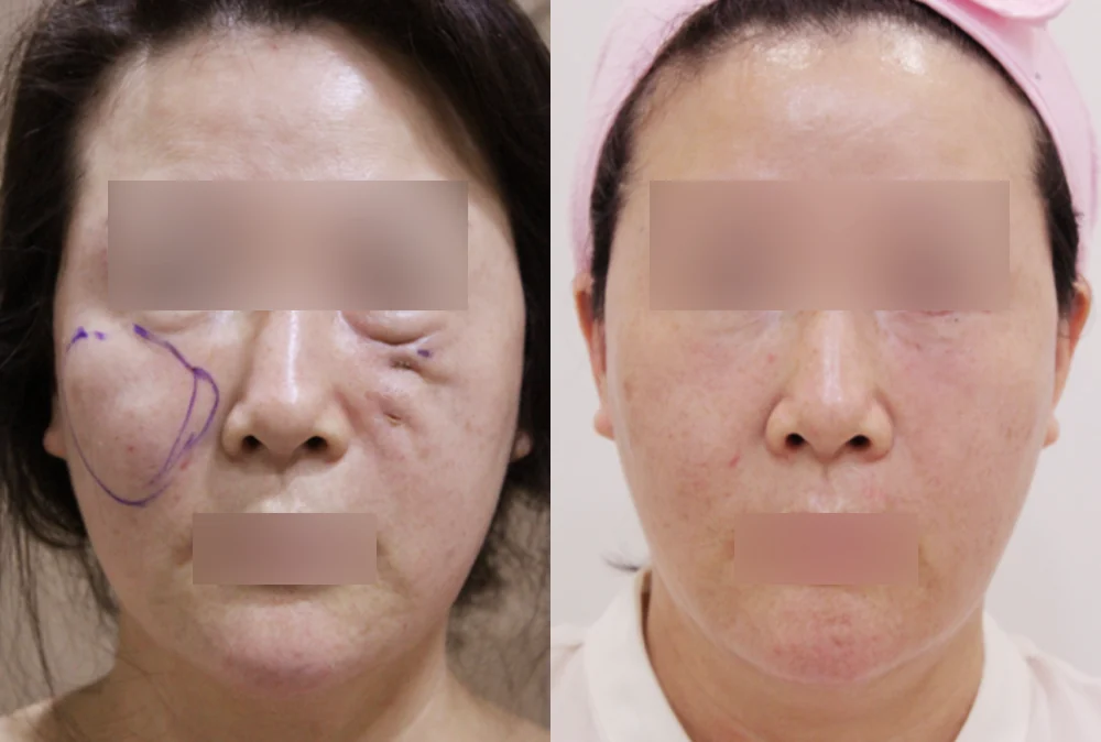
One cheek was raised in a large rounded bulge, and the other cheek had small scars and uneven marks
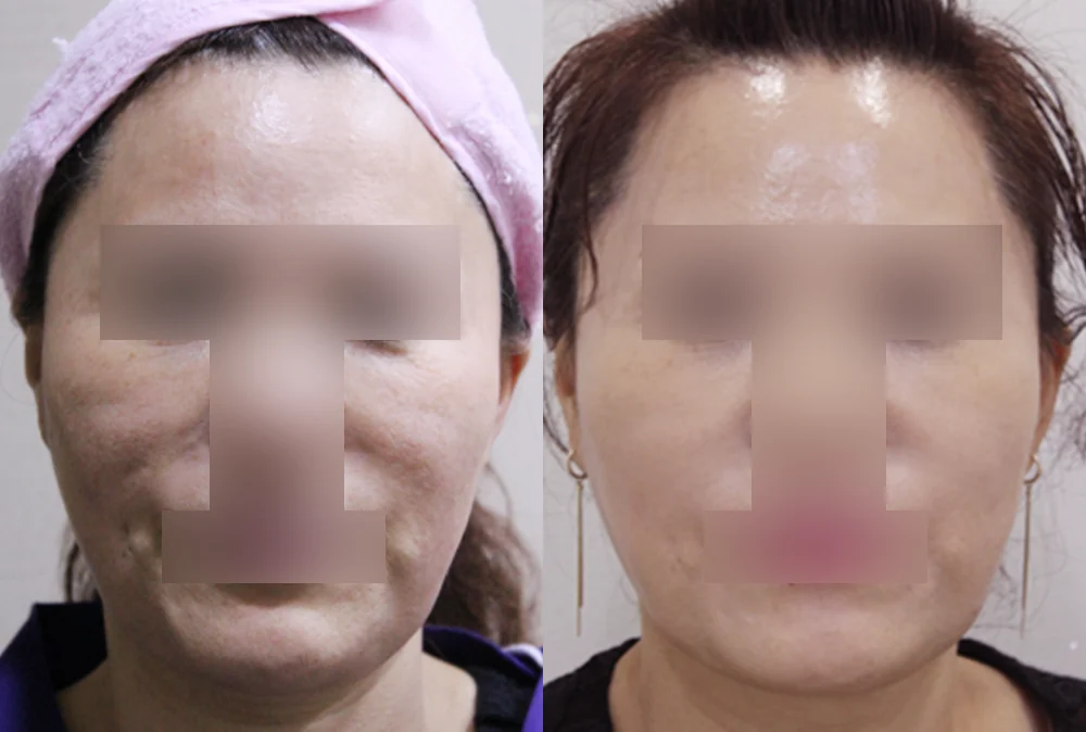
Lumps were raised on both cheeks beside the corners of the mouth, making the contour uneven
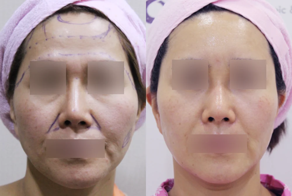
The contour was uneven across the forehead, cheeks and smile-line area (markings before surgery)
* Results vary from person to person. Side effects are possible, so please decide after a thorough consultation with a doctor.
Frequently asked questions
I have had no problems for over 30 years. Won't it stay that way?
It may. But how many people live their whole lives without trouble is hard to know, because most studies collect people who came to a hospital once a problem appeared. The material is still there, and granulomas can grow slowly years later.1 Having one ultrasound to record the present state makes any later change much easier to recognize.
I don't know whether it was silicone or paraffin. Can tests tell?
To some extent. Silicone has a characteristic appearance on ultrasound and MRI, and granulomas caused by silicone or paraffin can show rounded calcifications on CT.1 Even so, imaging cannot always settle exactly what the substance is. You can consult with us without knowing; tell us the year, the place and anything you were told at the time.
A screening mammogram or PET-CT showed something unusual. Could it be my old injections?
It could. There are reports of material injected into the face or neck moving down to the upper breast and showing as small white spots on a mammogram.4 On PET-CT, fillers can also light up strongly as a normal finding.1 Equally, the material can hide another condition. Tell the team who did the test about your injections and follow their advice on further tests. If the breast is involved, making sure a cancer is not missed comes first.5
If I decide to watch it, how often should it be checked?
There is no fixed interval. It depends on the amount and location of the material and on any change so far, so it is agreed with your doctor. If a lump grows or hardens, or the area becomes red and swollen, do not wait for the planned date; get it checked promptly.
Can old silicone or paraffin turn into cancer (a tumor)?
A foreign body granuloma is not a tumor; it is a lump of chronic inflammation that forms in response to material the body cannot dissolve.1 On scans, though, it can look like another disease, or hide one.1 If there is material in the breast, it is important not to miss a breast cancer that may be present at the same time.5 So a lump that is new or changing should be checked rather than simply put down to old injections.
What to prepare before your consultation
- When (even roughly), where, on which areas and how many times you were injected, and any substance name you were given
- Changes since then and when they began (swelling, lumps, redness, change in shape)
- Previous test results (ultrasound, MRI, mammography) and any abnormal blood results you were told about
- Other procedures on the same areas (fillers, threads, lasers, surgery)
- Front and side photos from before the injections and from now
Bonita Clinic is on the 3rd floor of 565 Gangnam-daero, Seocho-gu, Seoul, about 100 m on foot from Exit 8 of Nonhyeon Station (Line 7). You can read more on our forehead, chin and cheek revision page.
References (6)
Sources that support the explanations in this article. Study findings describe general trends and do not guarantee individual results.
- Mundada P, Kohler R, Boudabbous S, et al. Injectable facial fillers: imaging features, complications, and diagnostic pitfalls at MRI and PET CT. Insights Imaging. 2017;8(6):557-572.doi:10.1007/s13244-017-0575-0Imaging review (full text): filler classes (hyaluronic acid broken down within about a year vs liquid silicone that stays), paraffinoma and the silicone controversy, features of foreign body granulomas (months to years later, very slow growth, multiple or recurring, finger-like spread), late complications, and what ultrasound (snowstorm pattern hiding deeper tissue, operator-dependent), MRI (moved material, silicone-only sequence), CT (eggshell calcification) and PET-CT (normal uptake, may mask disease) can show
- Jang JW, Kang SY. Evaluation and management of facial granuloma caused by various injection materials. Arch Craniofac Surg. 2021;22(1):26-32.doi:10.7181/acfs.2020.00689Korean case series of 64 facial granulomas (full text): unknown illegal substances were the largest group (42.2%), unclear margins make complete removal difficult, factors that may trigger reactions, and in patients who already had symptoms, earlier surgery was linked to shorter treatment and higher satisfaction (retrospective)
- Harlim A, Andrew H, Harlim C. Novel Granulectomy Technique and Intralesional Triamcinolone for Liquid Injectable Silicone-Induced Chin Granulomas: A Long-Term Ambidirectional Cohort Study. J Clin Med. 2026;15(17):6506.doi:10.3390/jcm15176506Cohort study of 35 chin liquid-silicone granulomas in Indonesia (full text): granulomas often appear years after injection, silicone microdroplets spread so complete removal is nearly impossible, no treatment consensus; over a mean 137.9 weeks, recurrence was 44.4% (8/18) after excision alone vs 5.9% (1/17) with steroid injections afterwards
- Cho N, Kim EK, Oh KK, et al. Migrated foreign body granulomas on mammography after injection in the cervicofacial area. Clin Radiol. 2004;59(9):835-840.doi:10.1016/j.crad.2004.03.003Korean radiology case series (abstract): nine women with foreign material injected into the face and neck had migrated granulomas seen as multiple dense nodules under 5 mm in the upper inner breast on mammography; strong acoustic shadowing hid deeper neck structures on ultrasound
- Yen Kok KY, Tripathi S, Telisinghe P. Breast paraffinoma: An obsolete but still important condition - Experience with 60 cases and the literature review. Asian J Surg. 2023;46(1):321-327.doi:10.1016/j.asjsur.2022.04.002Case series and literature review of 232 breast paraffinoma patients (abstract): first symptoms were lump without pain 65.5%, painful lump 14.7%, ulcer 7.3% and others; mastectomy 53.4%, excision 17.7%, no further treatment 25%; excluding coexisting breast cancer is important
- Tachamo N, Donato A, Timilsina B, et al. Hypercalcemia associated with cosmetic injections: a systematic review. Eur J Endocrinol. 2018;178(4):425-430.doi:10.1530/EJE-17-0938Systematic review of 23 patients from 20 articles (abstract): hypercalcemia after cosmetic injections, mostly silicone (43.5%) in the buttocks (69.6%) or breasts (39.1%), appearing a mean 7.96 years after the procedure; kidney failure in 82.4%, two deaths; rare but delayed
About this article
This article separates general medical information from Bonita Clinic's own approach. Because the right decision depends on each person's condition, it is not a substitute for a consultation.