KOKorean
 ENEnglish
 JP日本語
 CN中文(简体)
 TW中文(繁體)


Dark Circles Under the Eyes: Causes, Types and Treatment in Seoul
Dark circles are not one condition but a shared name for several things that make the under-eye look dark, so the right treatment depends on which cause is present. The skin itself may hold extra brown pigment, vessels and muscle may show through thin skin as a bluish tint, or fat that has pushed forward may cast a shadow into the sunken groove below, and two or more of these often occur together. Pigment calls for treatment of the skin, while a shadow calls for treatment of the contour, so the causes need to be separated first. Lower eyelid fat repositioning is designed for the shadow made by a bulge and a hollow, and it does not remove pigment from the skin.
Curious about your own case? Ask us online →

Do dark circles have a single cause?
"Dark circles" is an everyday name for under-eye areas that look darker than the rest of the face. It is not a single diagnosis. Different causes can produce a very similar look in the mirror. A review written by Korean dermatologists (a paper that gathers and summarizes earlier research) notes that dark circles still have no clear definition, and that their causes have not been fully worked out.4
The causes this review lists are:4
- Extra pigment in the skin
- Shadows made by the tear trough (the natural groove that runs down and outward from the inner corner of the eye) and by under-eye fat that has pushed forward
- Shadows made by loose skin and wrinkles
- Thin, see-through skin over the orbicularis oculi (the ring muscle that closes the eye)
A dermatology study from Taiwan examined 65 people with dark circles using a Wood's lamp (an ultraviolet examination light) and ultrasound. It sorted them into four types: a brown pigmented type, a vascular type ranging from blue to purple, a structural type in which the contours of the face cast a shadow, and a mixed type with two or more of these together. The pigmented type made up 5%, the vascular type 14%, the structural type 3% and the mixed type 78%.1 Put simply, nearly eight in ten people had more than one cause at the same time.
At Singapore's National Skin Centre, researchers screened 1,000 consecutive dermatology patients and examined in detail 200 who had dark circles. Here the vascular type was the most common at 41.8%. Next came the constitutional type (pigment you are born with) at 38.6%, post-inflammatory hyperpigmentation (pigment left behind after skin inflammation) at 12%, and the shadow type at 11.4%. The vascular type was seen mainly in Chinese patients, and the constitutional type was most common in Indian and Malay patients.2
The two studies used different countries, patients and categories, so their numbers cannot be compared directly. Their shared message is clear, though. Dark circles often do not fit neatly into one type, and classifying them in detail helps in choosing a suitable treatment.1
The Korean review, for its part, considers the shadow type made by the tear trough, with or without bulging fat, the most common kind of clinically important dark circles.4 How the categories are drawn and who is studied can shift the proportions this much.
Which type are your dark circles closest to?
Two people can both say "my under-eyes look dark" and still have quite different colors and shapes. The four drawings below show the same person's under-eye, changed one type at a time.
Four types of dark circles by color and shadow
1A brown band, whatever the lighting
May be pigment in the skin itself; check whether the color stays when the skin is stretched
Skin treatment
2A bluish or violet tint shows through
Vessels and muscle may show under thin skin; puffiness is checked too
Skin and vessels
3A shadow sits under a bulge
May be the shadow of bulging fat and a sunken groove; the contour is checked
Structure
4Color and shadow appear together
The causes are separated and how much each contributes is assessed
Planned in parts
The card names are there to make the idea easier to follow. In real life the types often mix: brown pigment with a shadow, or a bluish tint with a bulge. The sections below look at why each type forms and what helps tell it apart.
Brown circles: the pigmented type
In the pigmented type, the under-eye skin itself holds more brown pigment (melanin), so it looks darker.1 The comprehensive review we draw on lists these causes:3
- Constitution and heredity
- Pigment left behind after atopic dermatitis or contact dermatitis (skin inflammation triggered by something that touches the skin, such as a cosmetic)
- Ultraviolet light, which makes under-eye pigment darker
Rubbing the eyes because they itch can also lead to post-inflammatory pigment.4 Some glaucoma eye drops, the prostaglandin analogues, can darken the skin around the eyes as well. With one of these drops, the darkening appeared most often three to six months after starting, and it reversed completely after the drop was stopped.3 These drops protect your sight, though. If the color bothers you, talk to your eye doctor first rather than stopping them yourself.
A key sign of the pigmented type is that the color stays when you gently stretch the skin.3 In dermatology, a Wood's lamp is sometimes used to estimate whether the pigment sits in the top layer of the skin (the epidermis) or deeper (the dermis). Pigment in the top layer looks more distinct under the lamp, while deeper pigment shows less contrast.3
Bluish or purple circles: the vascular type
The vascular type looks blue, pink or purple.1 The skin of the lower eyelid is thin and easy to see through. As a result, the orbicularis oculi muscle and the network of vessels within it can show through. In some people a fairly large vein also runs below the eye.4 This kind of darkness is described as a violet tint across the whole lower lid, often deeper toward the inner corner, and sometimes more noticeable during menstruation.3
It can come with puffiness around the eyes.3 If the darkness is worse when you wake up, or the morning after a salty meal, fluid build-up (edema) around the eyes may be playing a part.3 In children with allergic rhinitis, swollen nasal lining can slow the drainage of blood from the veins around the eyes, which can make the area look darker; these children often breathe through the mouth as well.4
When the skin of the vascular type is gently stretched, the dark area does not turn pale. The violet tint actually deepens, which is explained by thin eyelid skin or a rich supply of vessels.3 In the Singapore study the vascular type was the most common, and it was seen especially often in Chinese patients.2
A shadow under a bulge: the structural (shadow) type
In the structural type, the skin itself is not darker. Instead, the shape of the face casts a shadow that reads as darkness.1 Two structures usually create that shape.
The first is the tear trough, the natural groove that runs down and outward from the inner corner of the eye.4 The second is under-eye fat that has pushed forward. The Korean review explains that bulging fat does not directly create dark circles by itself. It makes the tear trough below look deeper, and that deepens the shadow.4 In the Taiwanese study, ultrasound confirmed that fat inside the orbit (the bony socket that holds the eye) had pushed forward in 14 people with under-eye bags.1
Loose skin and fine wrinkles, which increase with age, add further shadow.4 A key sign of this type is that the shadow fades, or disappears entirely, when the skin is stretched.3 The darkness also changes with the lighting.3 Our lower blepharoplasty guide shows how the direction of light changes the shadow, and our lower eyelid fat repositioning guide explains why fat pushes forward and why the area below it sinks.
The three causes sit at different depths. Pigment lies within the skin, visible vessels lie just beneath thin skin, and shadows come from the contour of the fat and the bony rim deeper down. That is why treatments for "dark circles" need to reach different places.
How can you tell the types apart at home?
There is a simple check you can try in front of a mirror: the stretch test described in the review.3
- In good light, place a fingertip near the outer corner of your eye and pull the skin very gently sideways. Do not pull hard or rub.
- Watch how the dark area changes.
- If the color stays the same, pigment in the skin is likely.
- If it does not turn pale and the violet deepens, vessels showing through are likely.
- If it fades or disappears, a shadow made by the contour is likely.
Changing the lighting helps too. A shadow changes a lot with the lighting,3 while the color of the skin itself looks much the same whichever way the light falls. Comparing morning and evening is also useful. If the area looks puffier and darker in the morning and improves during the day, swelling is probably part of the picture.3
These are clues, not a diagnosis. When several causes overlap, stretching makes part of the darkness fade while another part stays. As the Taiwanese study showed, this mixed picture is actually common.1 That is why the share of each cause is worked out at an examination.
What else is looked at during an examination?
Along with the overall appearance, an examination usually looks at:
- Color and location: whether it is brown, bluish or violet, and whether it is deeper at the inner corner
- Depth of pigment: dermatologists sometimes use a Wood's lamp to estimate whether the pigment sits in the top layer of the skin or deeper3
- Bulge and groove: whether under-eye fat has pushed forward and how deep the groove below is. The Taiwanese study used ultrasound to confirm bulging fat, and found that the tissue in front of the septum (the thin sheet that holds the fat back) was thicker in under-eyes that tended to be puffy.1 Ultrasound can also help tell a vascular cause from puffiness.3
- Skin and eyelid condition: how much loose skin and fine wrinkling there is, and whether the lower lid's support has weakened6
- History: atopic dermatitis or allergic rhinitis, eye-rubbing, current eye drops, and any previous filler, fat grafting or under-eye surgery
How does treatment change with the cause?
A systematic review (a study that gathers all relevant research using a set method) of 39 studies reached these conclusions:5
- For dark circles caused by volume loss, soft-tissue fillers and grafting of your own fat were the most effective.
- When marked skin looseness was the cause, eyelid surgery (blepharoplasty) was the most suitable option.
- For pigment, various creams and chemical peels (treatments that use a solution to remove the top layer of skin) were useful.
- Lasers were mildly to moderately helpful for both the vascular and the pigmented types.
A more recent systematic review of 50 studies published since 2010 points the same way. Fillers remained the foundation for volume loss. Lasers addressed pigment, vessels and skin looseness, and chemical peels and creams worked for color changes in the top layer of skin. Surgery was preferred when under-eye fat had pushed forward or the lower eyelid's support was weak.6
| Type | What is usually considered | Good to know |
|---|---|---|
| Pigmented | Sun protection, lightening creams, chemical peels, lasers | Hydroquinone, a typical lightening ingredient, is used as a medicine, usually takes five to seven months to show an effect and needs care with long-term use;3 creams can irritate the eye area4 |
| Vascular | Lasers, fat grafting to thicken thin skin | A long-pulsed 1,064 nm laser is sometimes used for visible veins;4 fat grafting is less likely than filler to look bluish or puffy, but swelling lasts longer and grafted fat is hard to remove4 |
| Shadow: mainly hollow | Filling with filler or your own fat | Effective for volume loss;5 too much volume can look puffy;4 rare cases of blocked blood vessels have been reported7 |
| Shadow: bulging fat | Surgery such as fat repositioning | Surgery is preferred when fat has pushed forward or lid support is weak6 |
| Shadow: loose skin | Lower blepharoplasty including skin removal | Most suitable when the skin is markedly loose5 |
| Mixed | Methods combined, cause by cause | No single method addresses every cause |
Each method also carries its own risks and limits. Lightening ingredients such as hydroquinone can cause irritation, itching and post-inflammatory pigmentation. Long-term use can lead to exogenous ochronosis, in which the treated skin turns blue-black.3 Filler placed too close to the surface can show through as a bluish tint, and too much can look puffy.4 Fat grafting is less prone to these problems, but the procedure takes longer, swelling can last a long time, and grafted fat is hard to remove.4
There is also a rare but more serious risk. A review of 98 reported cases of vision loss or vision change after filler or fat injections in the face found that the patient's own fat and hyaluronic acid filler were the most common materials, and that sight usually did not recover.7 Most reports involved the area between the eyebrows and the nose, but injections around the eyes also call for a doctor who knows the blood-vessel anatomy well.7
Both reviews are clear about a limitation: the quality of the evidence is not high. The first notes that high-quality studies are scarce, so its recommendations should be read selectively.5 The Korean review also points out that, although many treatments exist, there are no evidence-based studies to support their use.4 In plain terms, no treatment can promise to brighten everyone's under-eyes. Matching the method to the cause is the starting point.
When does fat repositioning fit, and when not?
Rather than throwing away the fat that has pushed forward, lower eyelid fat repositioning moves that fat into the sunken groove below to even out the contour. In other words, it is designed for the shadow created by a bulge and the groove beneath it. At Bonita Clinic, repositioning is the basic approach, and part of the fat is also removed when the condition calls for it. The way the fat is moved is explained in detail in our lower eyelid fat repositioning guide.
When a consultation is recommended
Our lower eyelid fat repositioning page recommends a consultation in these cases:
- Under-eye bulging and hollowing occur at the same time
- The under-eye hollow keeps progressing over time
- Under-eye lines remain where fat was removed, or the area has bulged again after surgery
- The under-eye surface is uneven after fat grafting or filler
If the shadow also fades when you gently stretch the skin, that is a further clue that the shadow is a large part of what you see.3
When other approaches come first
- Brown color with no bulge: this is pigment in the skin, so skin treatment comes first.5
- A bluish or violet tint with no bulge: vessels showing through are the main cause, so lasers or ways to thicken the thin skin are considered.4
- A hollow with no bulge: filling with filler or fat has been summarized as effective for volume loss, so it is considered alongside.5
- Marked loose skin with fine wrinkles: a lower blepharoplasty that also removes excess skin is considered.5
- Puffiness only in the morning that settles by midday: this may be edema, so causes such as allergies or salty food are looked at first.3 Eyelid swelling can also come from conditions elsewhere in the body or around the eye.3 If the swelling persists, affects only one side or comes with other symptoms, have the cause checked by an internist or eye doctor first.
Which direction comes first?
What stands out most under your eyes?
- A bulge with a shadowed groove belowConsider fat repositioningIf the skin is loose, add lower blepharoplasty
- A hollow without a bulgeFilling with filler or fat
- Brown color in any lightPigment treatment firstPlus sun protection
- A bluish or violet tintSkin-side options such as lasers or fillingIf puffy, look for the cause first
With a mixed type, the order and combination are planned in parts. For example, if a bulge and brown pigment appear together, surgery can reduce the shadow while the pigment in the skin remains. Knowing this beforehand helps set realistic expectations.
What helps in daily life?
- Sun protection: ultraviolet light darkens under-eye pigment. Broad-spectrum sunscreen and UV-blocking sunglasses are described as helpful. Because the eye area is delicate, the review advises caution with chemical sunscreens there.3
- Not rubbing: rubbing itchy eyes can cause post-inflammatory pigment. If you have atopic dermatitis or allergies, treating them comes first.34
- Watching for puffiness: puffiness that is worse in the morning or after salty food can be a sign of edema.3 If it lasts or affects one side only, have the cause checked.
- Sleep and stress: lack of sleep, stress, heavy drinking and smoking are often blamed, but the review notes this has not been clinically substantiated.3 Good sleep is good for your health, yet it is unlikely on its own to remove a structural shadow or pigment you were born with.
How we approach it at Bonita Clinic
Any surgery can cause side effects such as bleeding, infection or inflammation. A meta-analysis (a study that pools the results of many studies) of 33 studies and 4,671 patients who had under-eye fat grafting or repositioning found unsatisfactory correction or an uneven contour in 6.2% and a second operation in 2.4%.8 How the moved fat settles, and the limits of the operation, are explained in more detail in our lower eyelid fat repositioning guide. Recovery and results differ from person to person, and the actual treatment plan is decided after an in-person consultation.
Treatment options you can discuss in Seoul
Bonita Clinic has treated patients at the same location since opening in 1995, and Dr. Kwang-Seop Jung, a plastic surgeon board-certified in Brazil, sees patients himself. For dark circles, what you can discuss at consultation is the approach described on our treatment pages:
- An assessment of whether the darkness comes from color or from structure, and how much each contributes
- Lower eyelid fat repositioning through the conjunctiva for a bulge with a sunken groove below
- Lower blepharoplasty, which also tidies loose skin when skin looseness is a large part of the problem
- An assessment of an uneven under-eye surface after earlier fat removal, filler or fat grafting
Pigment and vessel color are a separate question from the contour. Which steps you need, and in what order, is decided only after an examination.
Why do the plan and cost differ from person to person?
Two people who both say "dark circles" can need very different plans. What shapes the plan, and therefore the cost, is which causes are present and how much needs to be done.
- Which causes are present: shadow, pigment, vessels
- Fat repositioning alone or with lower-lid skin removal
- Previous fat removal, filler or fat grafting
- Anesthesia: sedation
- Whether pigment or vessels need separate skin treatment
- How the bulge and hollow differ on each side
Final plan and quote after an in-person exam
A final estimate is given only after an in-person examination, once the causes and the extent of treatment are clear. Photos help narrow down the options, but the plan is settled at the examination.
What can photos show, and what needs an in-person exam?
Photos are a good place to start, because color and contour show up well in pictures. They cannot show how the skin reacts when stretched, how much fat lies behind a bulge or how firmly the lower lid is supported.
Photos can show
- Brown or bluish color in daylight
- Where it is darkest: inner corner or whole lid
- A bulge and the groove below it
- How the shadow changes under ceiling light
- Morning versus evening puffiness
Needs an in-person exam
- How the color reacts when the skin is stretched
- Whether color or contour is the main cause
- How much fat bulges, and where
- Lower-lid support and skin looseness
- Remaining filler or grafted fat
Helpful photos: your under-eyes from the front in daylight without makeup, a photo under ordinary ceiling light, and both 45-degree views. If puffiness changes through the day, a morning and an evening photo help. Please also mention any previous filler, fat grafting or under-eye surgery. You can send us a message with your photos and a short description of what bothers you most: the color, the bulge or the hollow.
Planning your visit and recovery
What is confirmed in advance is the treatment information above: fat repositioning under sedation, same-day discharge and no stitches to remove. How long you should stay in Seoul and how many follow-up visits you need depend on the extent of treatment, for example whether lower-lid skin is removed at the same time. These are planned together with you at consultation.
Swelling and bruising are expected after eyelid surgery, and the under-eye contour settles gradually as the swelling goes down. If you have an important event or a fixed return date, tell us at consultation so the plan can take it into account. After you return home, contact a doctor promptly if pain, redness, swelling or bleeding increases instead of settling, or if your vision changes. Dated photos taken in the same light make it easier for any doctor to follow your progress. Pigment or vessel color that remains after surgery is a separate matter; ask at consultation how it can be followed up once you are home.
Real treatment cases
These cases are from our lower eyelid fat repositioning page. All of them show a shadow under an under-eye bulge, and results vary from person to person.
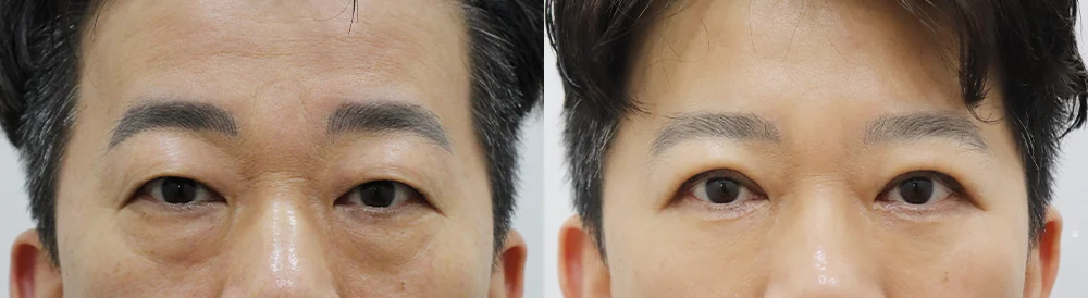
A bulging under-eye with a shadow below (front view)
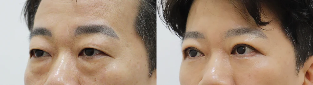
The same case seen at an angle
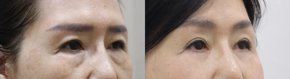
An angled view showing an under-eye bulge with a shadowed groove below
* Results vary from person to person. Side effects are possible, so please decide after a thorough consultation with a doctor.
Frequently asked questions
Will more sleep make my dark circles go away?
Lack of sleep and stress are often blamed, but the review notes that this has not been clinically substantiated.3 Puffiness that is worse in the morning and then improves may change with daily habits. Pigment you were born with, or a shadow made by bulging fat, is unlikely to disappear with sleep alone. If the darkness looks much the same every day, it is worth finding out the cause.
Can eye creams or brightening cosmetics help?
Cosmetics and medicines are not the same thing. If pigment in the skin is the cause, a lightening cream may help.5 Hydroquinone, a typical lightening ingredient, is treated as a medicine rather than an ordinary cosmetic ingredient in many countries, so it should be used for a set period under a doctor's guidance. It usually shows an effect only after five to seven months, so the review suggests using it for at least three months.3 Irritation, itching and post-inflammatory pigmentation can occur, and long-term use has been linked to exogenous ochronosis, a blue-black darkening of the skin.3 If the darkness is a shadow made by the contour, a cream will not change the shape.
Will fat repositioning also lighten the color of my dark circles?
The shadow made by the contour can lessen once fat is moved into the groove. But this operation does not remove pigment from the skin. If brown pigment or visible vessels are also present, that color can remain, and skin treatment may be considered separately. This is why the share of each cause is checked before surgery.
My under-eyes look bluish after filler. Why?
When filler sits too close to the surface, it can show through as a bluish tint, known as the Tyndall effect. For this reason the Korean review recommends placing under-eye filler in the deep layer beneath the muscle, and warns that too much volume can make the area look puffy.4 If you already notice this change, the position and amount of the remaining filler should be checked first. See our guide to under-eye, glabella and nose filler removal for more.
Is one laser session enough for dark circles?
In a systematic review, lasers were mildly to moderately helpful for the pigmented and vascular types.5 In a study of pigmented dark circles, 15 of 18 patients who used a laser together with a lightening ointment had good or excellent results after three to four sessions.3 In other words, several sessions are common, and how much it helps varies from person to person. A shadow created by bulging fat is unlikely to change with lasers alone, which is why surgery is considered first in that case.6
What to prepare before your consultation
- Front-view photos of your under-eyes in daylight, plus one under ceiling light
- Whether the darkness or puffiness differs between morning and evening
- Atopic dermatitis or allergic rhinitis, eye-rubbing habits and any eye drops you use
- Previous filler, fat grafting, laser or under-eye surgery, with approximate dates
- What bothers you most: the color, the bulge or the hollow
Bonita Clinic is on the 3rd floor of 565 Gangnam-daero, Seocho-gu, Seoul, about 100 m on foot from Exit 8 of Nonhyeon Station (Line 7). You can read more on our Lower Eyelid Fat Repositioning page and Lower Blepharoplasty page. The difference between removing and moving fat is covered in our lower eyelid fat repositioning guide, and surgery when loose skin is also present in our lower blepharoplasty guide.
References (8)
Sources that support the explanations in this article. Study findings describe general trends and do not guarantee individual results.
- Huang YL, Chang SL, Ma L, et al. Clinical analysis and classification of dark eye circle. Int J Dermatol. 2014;53(2):164-170.doi:10.1111/j.1365-4632.2012.05701.xDiagnostic study of 65 dermatology patients in Taiwan (abstract): Wood's lamp and ultrasound used to classify dark circles as pigmented (brown), vascular (blue to purple), structural or mixed, at 5%, 14%, 3% and 78%; 33 puffy cases had thicker pre-septal tissue than 20 controls, and 14 with under-eye bags had protruding fat on ultrasound; detailed classification helps choose treatment
- Ranu H, Thng S, Goh BK, et al. Periorbital hyperpigmentation in Asians: an epidemiologic study and a proposed classification. Dermatol Surg. 2011;37(9):1297-1303.doi:10.1111/j.1524-4725.2011.02065.xEpidemiologic study at Singapore's National Skin Centre (abstract): of 1,000 consecutive patients, 200 with dark circles were examined; vascular 41.8%, constitutional 38.6%, post-inflammatory hyperpigmentation 12%, shadow 11.4%; the vascular type was seen mainly in Chinese patients and the constitutional type in Indian and Malay patients (a cross-sectional epidemiologic study); the four percentages add up to 103.8% as reported
- Sarkar R, Ranjan R, Garg S, et al. Periorbital Hyperpigmentation: A Comprehensive Review. J Clin Aesthet Dermatol. 2016;9(1):49-55.Comprehensive review (full text): causes incl. heredity, post-dermatitis pigment, edema (systemic or local), vessels, laxity and tear-trough shadow; vascular type with or without puffiness; stretch test (pigment persists, shadow fades, violet deepens); Wood's lamp for epidermal vs dermal pigment; ultrasound; sunscreen; sleep and stress not clinically substantiated; prostaglandin eye-drop pigmentation, reversible; hydroquinone effect after 5 to 7 months, side effects incl. irritation and exogenous ochronosis; ruby laser with bleaching good in 15 of 18
- Park KY, Kwon HJ, Youn CS, et al. Treatments of Infra-Orbital Dark Circles by Various Etiologies. Ann Dermatol. 2018;30(5):522-528.doi:10.5021/ad.2018.30.5.522Korean dermatology review (full text): no clear definition; causes include pigment, tear-trough and fat-herniation shadow, laxity, thin skin and large veins; fat herniation deepens the tear trough; tear-trough type most common clinically; allergic rhinitis with mouth breathing and venous stasis from swollen nasal lining; deep filler to avoid Tyndall effect and puffiness; fat grafting less puffy than filler but slower, longer edema, hard to remove; long-pulsed 1,064 nm laser for veins; little evidence-based support
- Michelle L, Pouldar Foulad D, Ekelem C, et al. Treatments of Periorbital Hyperpigmentation: A Systematic Review. Dermatol Surg. 2021;47(1):70-74.doi:10.1097/DSS.0000000000002484Systematic review of 39 studies (abstract): fillers and autologous fat grafting most effective for volume loss, blepharoplasty for marked skin laxity, creams and peels for pigment, lasers mildly to moderately helpful for vascular and pigment types; high-quality evidence is scarce, so recommendations should be read selectively
- Beer J, Boghosian T, Sherif R, et al. What's New With Under Eye Treatment: A Multispecialty Systematic Review of Recent Under Eye Treatments. Dermatol Surg. 2026;52(2):155-163.doi:10.1097/DSS.0000000000004964Multispecialty systematic review of 50 studies since 2010 (abstract): fillers are foundational for volume loss, lasers address pigment, vessels and laxity, peels and topicals suit superficial color change, and surgery is preferred for herniated fat and poor lower-lid support; fat, skin and lid support guide individual choice
- Beleznay K, Carruthers JDA, Humphrey S, et al. Avoiding and Treating Blindness From Fillers: A Review of the World Literature. Dermatol Surg. 2015;41(10):1097-1117.doi:10.1097/DSS.0000000000000486Literature review of 98 cases of vision change after filler injection (abstract): high-risk sites were the glabella 38.8%, nose 25.5%, nasolabial fold 13.3% and forehead 12.2%; autologous fat (47.9%) and hyaluronic acid (23.5%) were the most common materials; most vision loss did not recover; rare, but injectors need firm knowledge of vascular anatomy
- Xu Z, Zheng F, Zhao X, et al. Complications of Fat Grafting and Repositioning for Correction of Lower Eyelid Pouch With Tear Trough Deformity or Lid-Cheek Junction: A Systematic Review. Ann Plast Surg. 2024;93(3):e9-e25.doi:10.1097/SAP.0000000000004022Systematic review and meta-analysis of 33 studies and 4,671 patients (abstract): after under-eye fat grafting and repositioning, total complications 11.2%, unsatisfactory correction or contour irregularity 6.2%, hematoma, swelling, bruising or oozing 6.2%, reoperation 2.4%
About this article
This article separates general medical information from Bonita Clinic's own approach. Because the right decision depends on each person's condition, it is not a substitute for a consultation.